
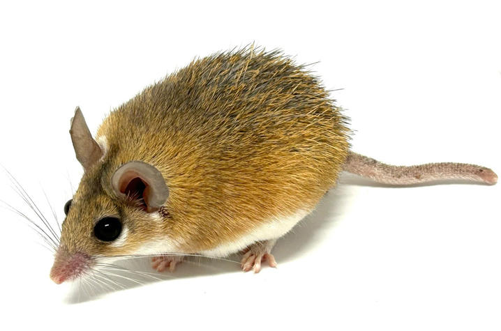
LEXINGTON, Ky. (Jan. 25, 2024) — A team of researchers at the University of Kentucky and Cincinnati Children’s Hospital is exploring the science behind how spiny mice can regenerate lost tissue and using what they learn to trigger regeneration in other types of mice. These advances may one day benefit into humans.
Whereas adult laboratory mice heal injuries with scar tissue, spiny mice can regrow lost skin and regenerate musculoskeletal tissues in their body.
Ashley W. Seifert, Ph.D., an associate professor in the Department of Biology in the UK College of Arts and Sciences, and his research group have pioneered using spiny mice and other animal models to understand how complex tissue can regenerate, bridging regenerative biology and medicine.
In April 2023, Science Advances published a study from Seifert’s group focused on the cellular response to injury during tissue healing. The paper showed how ERK signaling acts as a key switch balancing the healing response, and it demonstrated that increasing and maintaining this type of signaling in laboratory mice could trigger a regenerative response instead of scarring.
Their latest study, published in Developmental Cell, provides new insight into how specific immune cells respond to injury and direct tissue regeneration.
The team of scientists looked at a specific type of immune cell known as macrophages, which play a crucial role in regulating the inflammatory response by defending injured tissue against pathogens while simultaneously promoting tissue repair in both spiny mice and lab mice.
“In our study, we used a dual-species system to dissect macrophage phenotypes between two modes of healing: tissue regeneration or scar tissue formation,” said Seifert. “While it’s still unclear the extent to which macrophages ultimately control different healing trajectories, we observed subtle and distinct macrophage signatures that aligned specifically with regeneration.”
Using an identical injury in both kinds of rodents, the researchers studied signals released by macrophages that communicate to nearby cells, telling them to rebuild all the tissues that are missing. They also noted the characteristics, or phenotypes, of the cells during those processes.
“Our study shows that spiny mouse macrophages release distinct proteins that are partially responsible for the reformation of specialized tissues at the site of injury and for protecting cells from stress,” Seifert said. “Overall, our experiments point towards macrophages establishing a tissue microenvironment conducive for regeneration.”
Researchers examined and compared macrophages from the bone marrow of both types of mice. Then they used RNA sequencing to identify proteins these cells secrete. (RNA, or ribonucleic acid, is a molecule that carries genetic instructions from DNA to create proteins.)
Researchers found that the macrophages from bone marrow responded differently on their own to interferon gamma, a protein produced by cells in response to infection, and lipopolysaccharide, a molecule found in some bacteria that stimulates the immune system.
“In response to inflammatory cues, macrophages in spiny mice release unique communicating proteins that promote the growth of new blood vessels, lymphatic vessels, anti-inflammatory activities and tissue rebuilding that positively contribute to regenerative healing,” said Seifert.
The UK team found that those cells maintain their uniqueness throughout scar tissue formation or regeneration. Scientists studied samples at five different time points during healing to better understand the timeline for how macrophages do their work.
A gene analysis showed researchers which types of molecules produced by the cells were most active in each type of mice and with which functions those were most associated.
“In our work to answer more scientific questions about spiny mice, we found that a specific type of protein — vascular endothelial growth factor c or VEGFC — is uniquely secreted by spiny mouse macrophages during regeneration,” said Seifert. “It also plays a multifunctional role, encouraging the growth of new blood and lymphatic vessels.”
“Through specific antibody blocking of VEGFC protein in the ears of spiny mice, we observed notable changes in the formation of blood vessels, lymph vessels and cell division. This also led to decreases in new hair follicle formation and increases in inflammation, ultimately disrupting the process of tissue regeneration,” said Ajoy Aloysius, Ph.D., co-first author of the study and a postdoctoral scholar in biology. “Additionally, we speculate that enhancing the secretion of VEGFC and other factors by macrophages during fibrotic wound healing may contribute to a regenerative outcome in tissue healing, however further experiments are required to confirm this idea.”
The study ultimately suggests that macrophages found in specific tissues throughout the body help direct and regulate the cellular repair program. Importantly, the results of this study show that the type of macrophage matters and suggest that altering what one type of macrophage secretes could alter how tissue repairs itself.
“This study aims to unlock the body’s natural potential to regenerate after traumatic injury,” said Jennifer Simkin, Ph.D., one of the study’s first authors and an assistant professor of orthopaedic surgery at the Louisiana State University Health Sciences Center. “To do this, we study animals that can regenerate multiple tissues (hair, cartilage, muscle and skin) after injury to find the cellular and molecular signals necessary for better healing. Ultimately, we hope these findings pave the way for novel therapies to enhance wound healing.”
Researchers say further study is needed to better understand and define some of this cellular crosstalk during the regeneration process.
This team of researchers represents the UK Departments of Biology and Physiology, the UK Spinal Cord and Brain Injury Research Center, Louisiana State University Health-New Orleans, the University of Cincinnati Children’s Hospital Medical Center and the European Bioinformatics Institute.
Research reported in this publication was supported by the National Institute of Arthritis and Musculoskeletal and Skin Diseases of the National Institutes of Health under Award Number R01AR070313, the National Institute of Dental and Craniofacial Research of the National Institutes of Health under Award Number R21DE028070, the National Center for Advancing Translational Sciences of the National Institutes of Health under Award Number UL1TR001998, and the National Cancer Institute of the National Institutes of Health under Award Number P30CA177558. The content is solely the responsibility of the authors and does not necessarily represent the official views of the National Institutes of Health.
As the state’s flagship, land-grant institution, the University of Kentucky exists to advance the Commonwealth. We do that by preparing the next generation of leaders — placing students at the heart of everything we do — and transforming the lives of Kentuckians through education, research and creative work, service and health care. We pride ourselves on being a catalyst for breakthroughs and a force for healing, a place where ingenuity unfolds. It's all made possible by our people — visionaries, disruptors and pioneers — who make up 200 academic programs, a $476.5 million research and development enterprise and a world-class medical center, all on one campus.
In 2022, UK was ranked by Forbes as one of the “Best Employers for New Grads” and named a “Diversity Champion” by INSIGHT into Diversity, a testament to our commitment to advance Kentucky and create a community of belonging for everyone. While our mission looks different in many ways than it did in 1865, the vision of service to our Commonwealth and the world remains the same. We are the University for Kentucky.