Biology Ribble Endowment Seminar

"Glial Control of C. elgans Behavior"

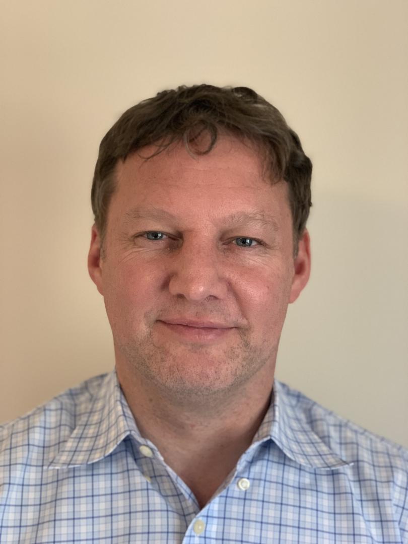
Shaham

Dr. Shai Shaham | Shaham Lab

Bio:
Glial cells are major components of nervous systems, and are in a position to influence nearly every
step of neural information transfer and processing. To understand if and how glia control nervous system
functions, the Shaham lab developed the nematode C. elegans as a unique setting to probe glia-neuron
interactions; demonstrating that glia in this animal can be interrogated without perturbing neuronal viabilityan
important experimental advantage. Using molecular, cell-biological, and physiological tools, developed in part by the Shaham lab group, the lab identified multiple mechanisms by which glia influence nervous system development and function. The Shaham lab showed that the four CEPsh glia of C. elegans are astrocyte-like glia that play central roles in modulating locomotory behavior. The Shaham lab also investigated glia that ensheath sensory-neuron receptive endings, and identified novel signaling interactions with these neurons that control an animal’s response to environmental stimuli. Dr. Shaham will describe recent published and unpublished findings that support the notion that glia play active and critical roles in defining behaviorally consequential activity set points in the nervous system. The lab hypothesizes that many of the rules we describe are conserved across animals, and Dr. Shaham will discuss evidence that
supports this idea.

Shai Shaham received his A.B. degree in biochemistry from Columbia University in 1989. In 1995, he graduated from the Massachusetts Institute of Technology with a Ph.D. in biology. After postdoctoral studies at the University of California, San Francisco, Shaham joined Rockefeller as assistant professor in 2001. He was named associate professor in 2007 and professor in 2012. Shaham was named a Sidney Kimmel Foundation for Cancer Research Scholar and a Rita Allen Foundation Scholar. He has received an Irma T. Hirschl/Monique Weill-Caulier Trust Research Award, a Masin Young Investigator Award from the Breast Cancer Alliance, a Klingenstein Fellowship, The Rockefeller University Distinguished Teaching Award, a Blavatnik Award, and a NINDS Outstanding Investigator Award.

Abstract:
Glial cells are major components of nervous systems and are in a position to influence nearly every step of neural information transfer and processing. To understand if and how glia control nervous system functions, we developed the nematode C. elegans as a unique setting to probe glia-neuron interactions; demonstrating that glia in this animal can be interrogated without perturbing neuronal viability- an important experimental advantage. Using molecular, cell-biological, and physiological tools, developed in part by our group, we identified multiple mechanisms by which glia influence nervous-system development and function. We showed that the four CEPsh glia of C. elegans are astrocyte-like glia that play central roles in modulating locomotory behavior. We also investigated glia that ensheath sensory-neuron receptive endings, and identified novel signaling interactions with these neurons that control an animal’s response to environmental stimuli. Our studies support the notion that glia play active and critical roles in defining behaviorally consequential activity set points in the nervous system. We hypothesize that many of the rules we describe are conserved across animals, and will provide evidence that supports this idea.

Watch the seminar here!

Date:
Location:
THM 116

"Tapeworm Tails: Signals that drive Stem Cell-dependent Regeneration in Hymenolepis diminuta"

RozarioDr. Tania Rozario | Rozario Lab

Bio:
Tania Rozario got her PhD from the University of Virginia studying embryonic development. During her postdoc she joined Phil Newmark's lab (Morgridge Institute for Research, WI) to study planarian regeneration but pivoted toward their parasitic cousins- tapeworms. Her work (re)established the rat tapeworm, Hymenolepis diminuta, as a non-traditional model to explore the molecular mechanisms that govern how tapeworms grow, regenerate, and reproduce at prolific rates. In 2021, she established her independent lab at the University of Georgia where her work understanding extrinsic and intrinsic signals that regulate tapeworm stem cells continues.

Abstract:
Tapeworms grow at rates that rival all metazoan tissues, including during embryonic and neoplastic growth. The rat tapeworm, Hymenolepis diminuta, produces up to 2,200 proglottids (segments), increasing in length up to 3,400 fold, and weight up to 1.8 million fold within the first 15 days of infection. Tapeworms can also regenerate: they shed large parts of their body, releasing their embryos to continue their life cycle, yet are able to continuously replenish proglottids and maintain an equilibrium length. Despite their impressive feats of growth, regeneration-competence is limited to one anatomical region- the neck. Using transcriptomics and RNA interference we have functionally validated the first molecular regulators of tapeworm regeneration and demonstrated that regeneration is dependent on a large population of poorly understood stem cells. Uncovering neck-exclusive stem cell subpopulations that can explain regionally restricted regeneration has remained elusive. Instead, we find that lethally irradiated tapeworms can be rescued from death when cells from both regeneration-competent and regeneration-incompetent regions are transplanted into the neck, suggesting that extrinsic signals at the neck are crucial for regeneration. In pursuit of such signals, we have discovered that the head has an organizer-like function. The head both maintains neck identity and regulates stem cell proliferation by establishing polarized expression patterns of Wnt signaling components like sfrp and beta-catenin. Our work is beginning to elucidate how the head and neck provide a rich signaling environment that enables region-specific regeneration in tapeworms.


Date:
Location:
THM 116

"Bee Biodiversity and Natural History Collections"

Seltmann

Dr. Katja Seltmann 

Bio:
Katja Seltmann is the Director of the Cheadle Center for Biodiversity and Ecological Restoration at the University of California, Santa Barbara. The Cheadle Center manages 400 acres of restored habitat in coastal central California and maintains a natural history collection of over half a million specimens. Her research blends data science, digitized collections, and media arts to understand insect biodiversity, conservation, and evolution. Katja is currently leading the "Extending Anthophila Research Through Image and Trait Digitization" (Big-Bee) project, funded by the U.S. National Science Foundation. This multi-year initiative focuses on digitizing bee collections by capturing high-resolution images of bee specimens and creating detailed datasets of their traits. The project involves collaboration with thirteen U.S. institutions and government agencies, aiming to enhance research capabilities and support bee biodiversity conservation efforts.

Abstract:
Functional traits of bees, such as pilosity (hairiness), wing patterns, and dietary preferences, are important for understanding their ecology and evolution. These traits influence pollen collection, pollination efficiency, temperature regulation, and resilience to environmental changes. In this seminar, I will share our work at UC Santa Barbara's Cheadle Center for Biodiversity and Ecological Restoration, where we utilize computer vision and machine learning to analyze high-resolution bee images and large specimen datasets from natural history collections. Our methods offer innovative ways to explore bee biodiversity, including findings that climate and evolutionary history may influence bee hair patterns, that population variations can be detected through wing venation analysis, and that the pollen diet of bees can be predicted based on range size and other factors. Overall, our research provides deeper insights into bee biology and trait evolution and shows potential for improving bee health and conservation monitoring by identifying traits related to resilience and stress.

Beautiful bee


Date:
Location:
THM 116

"Genetic and Genomic Mechanisms Underlying the Convergent Evolution of Pollination Syndromes in the Neotropical Radiation of Costus L. (Costaceae)"

Specht

Dr. Chelsea Specht | Specht Lab

Bio:
Dr. Chelsea Specht is the Barbara McClintock Professor of Plant Biology and Associate Director for Faculty Development, Equity, and Inclusion in the School of Integrative Plant Science and serves as the elected Associate Dean of Faculty for Cornell University.   She is a faculty member in the graduate fields of Plant Biology and Ecology and Evolutionary Biology and a faculty fellow of the Atkinson Center for a Sustainable Future.  She is also a member of the L.H. Bailey Hortorium and affiliated with the Cornell University Herbarium. 

In the Specht Lab we work together to investigate the evolution and diversification of Plant Form and Function. We use traditional morphological and developmental techniques combined with molecular genetics, comparative genomics and evolutionary biology to study the natural diversity of plants and to help better understand the forces creating and sustaining this diversity.  Our research incorporates elements of systematics, developmental genetics and molecular evolution to study the patterns and processes associated with plant speciation and diversification.  We take advantage of living and preserved collections to advance our research in plant systematics, biogeography, and developmental evolution. 

Abstract:
Fifty years since Dr. Paul J.M. Maas published his first monograph of the New World Costoideae, we continue to struggle with species boundaries and evolutionary relationships within this charasmatic lineage of tropical monocotyledonous plants.  In fact, the more we explore and discover the more questions emerge about the dynamics, patterns, and processes leading to speciation and diversification across the Neotropical Costaceae.  In this seminar, I will discuss the recent monographic revision and its critical role in establishing a framework for evolutionary and ecological studies of the Neotropical Costus lineage within a phylogenetic context.  The tempo and mode of speciation events are correlated with morphological changes that influence organismal interactions, including pollination and herbivory.  Ecologic, morphologic, and biogeographic conditions that appear to promote hybridization and result in the potential for hybrid speciation are discussed across the genus, and implications for developing a stable taxonomy – and whether or not that is even possible or desirable – will be discussed.

Date:
Location:
THM 116

"The Dual Role of Parental Conflict in Speciation: Lessons from Mimulus""

Coughlan_Selfie

Dr. Jennifer Coughlan | Coughlan Lab

Bio:
See attached CV here.

Abstract:
Determining what factors generate biodiversity is a central question in evolutionary biology. Despite its long history of study, we are only beginning to understand the evolutionary drivers of reproductive barriers between species, including reproductive barriers that manifest as sterile or dead hybrids. An intriguing hypothesis is that intragenomic conflicts- or selfish evolution- can drive the evolution of alleles that cause hybrid sterility/inviability. One such source of conflict is conflict between parents over resource allocation to offspring. Under parental conflict, multiple paternity drives the evolution of paternally derived, resource-acquiring alleles, and maternally derived alleles that distribute resources equally among offspring. In hybrids, mismatches between these parent-of-origin effect alleles can cause inappropriate development of placenta or endosperm, and subsequently embryo death. Here, I test the role of parental conflict in generating one of the most common intrinsic barriers in seed angiosperms- hybrid seed inviability-using members of the evolutionary and ecological model system; the Mimulus guttatus species complex. I show that hybrid seed inviability has evolved rapidly and repeatedly in this group, and patterns of HSI conform to the predictions of parental conflict. Additionally, genetic mapping suggests that hybrid seed inviability is conferred by nuclear, parent-of-origin effect loci (i.e. loci that affect the probability of death only if maternally or paternally derived). Lastly, using a series of natural surveys and mixed pollination crosses, I find that species with different histories of parental conflict frequently co-occur and hybridize, and hybridization between species with differing histories of parental conflict can indirectly influence growth in intraspecific seeds. Overall, this work highlights a dual role of parental conflict in the speciation process; both in the origin of reproductive isolation, but also in the dynamics and outcomes of hybridization in nature.

Watch the seminar here!
 

Date:
Location:
THM 116

"The Developmental Basis of Phenotypic Diversity"

Dr. Ricardo Mallarino | Mallarino Lab

Bio:
Ricardo Mallarino is an Assistant Professor of Molecular Biology at Princeton University. Originally from Bogota, Colombia, he graduated with a B.S. in Biology from Universidad de los Andes. He completed his graduate studies at Harvard in Organismic and Evolutionary Biology in 2011, working with Arhat Abzhanov on developmental mechanisms underlying beak shape diversity in Darwin’s finches and their close relatives. After completing his PhD. he joined Hopi Hoekstra’s lab at Harvard, where he established a new model species and developed tools for studying the molecular basis of pigment pattern formation in mammals. Research in the Mallarino lab focuses on understanding the genetic and developmental mechanisms by which form and structure are regulated during vertebrate embryogenesis and elucidating how these processes get modified during evolutionary time to produce phenotypic diversity.

Abstract:
The evolution of metazoan organisms over millions of years has led to remarkable complexity of form and function. While biologists have long studied the ultimate causes of biological diversity (i.e., why it originates), the proximate mechanisms underlying its emergence (i.e., how it arises) remain largely unknown. The goal of my lab is to uncover the genetic and developmental mechanisms underlying the establishment of phenotypic traits and to understand how these mechanisms have evolved to generate diversity across species. We achieve this by harnessing naturally evolved phenotypic variation in ‘non-traditional’ species and integrating multiple disciplines, including developmental biology, computational biology, and evolutionary genetics. For the past six years, our research has focused primarily on patterning and evolution of novelty in mammalian skin. In this talk, I’ll describe how my lab has developed new model systems to study two distinct spatially patterned phenomena during skin development - stripe pattern formation in rodents and gliding membrane formation in marsupials. Through the use of experimental embryology, transcriptomics, comparative genomics, and functional genetics, our work has yielded insights into the mechanisms by which phenotypic novelty is generated at the molecular level.

Watch the seminar here!

Date:
Location:
THM 116

"Phylodynamic and Comparative Approaches for Reconstructing Major Evolutionary Transitions in Deep Time"

Dr. Tiago Simões | Simões Lab

Bio:
Dr. Tiago Simões started his career in his home city (Rio de Janeiro, Brazil), where he obtained his BSc and MSc in Biological Sciences- Zoology at the Federal University of Rio de Janeiro and the National Museum of Brazil. He obtained his PhD at the University of Alberta, Canada, in 2018 working with Dr. Michael Caldwell. Between 2019 and 2023 he was a Postdoctoral Fellow at the Museum of Comparative Zoology & Dpt. Organismic and Evolutionary Biology, Harvard University, working with Dr. Stephanie Pierce, and since 2022 a Research Associate in the Division of Vertebrate Zoology at the American Museum of Natural History. Since 2024, he has been an Assistant Professor in the Dpt. Ecology and Evolutionary Biology at Princeton University.

Dr. Simões’s research integrates data from living and extinct species, as well as morphological and genomic data, to investigate deep time problems in vertebrate evolution, with a special focus on the origin and early evolution of lizards and snakes. He has created several new morphological and total-evidence datasets employing state-of-the-art techniques in Bayesian phylogenetics and phylodynamics that helped bridging gaps between morphological and molecular hypothesis of reptile evolution. These studies, along with new technical advances in phylogenetics have been published in several peer-reviewed scientific articles creating, including in Nature, Nature Ecology & Evolution, and Science Advances

:
The history of life on Earth is marked by complex interactions between species genomes and phenotypes across constantly changing environments. Therefore, it is necessary to investigate these interactions across deep evolutionary time to understand the processes responsible for the construction of both past and modern biological diversity. However, this line of research has historically faced several logistic and methodological limitations, such as the lack of quantitative methods for combining various data types sampled across vastly different organismal and temporal dimensions. Fortunately, the past decade has been testimony to several advances in Bayesian evolutionary analyses that have fostered the integration of data types towards more sophisticated inferences of evolutionary trees and macroevolutionary dynamics. Here, I will illustrate how I have used and expanded this class of techniques to integrate molecular and phenotypic data from living and fossil species to understand the patterns and processes operating across major evolutionary transitions in vertebrates, with a special focus on reptiles. These results have overhauled the structure of key areas of the reptile tree of life, including the origin of lizards and turtles, the interplay between phenotypic and molecular innovations during evolutionary transitions, and how these events have been impacted by climate change across deep time. I conclude by highlighting how a new omics era, integrating whole genomes and phenomes, can conciliate historical challenges in understanding organismal evolution and the interplay between genomes and phenotypes with their surrounding environments across broad taxonomic and time scales.

Watch the seminar here!

Date:
Location:
THM 116

"Evolution of Floral Disparity through Integration of Fossil and Extant Morphological Diversity"

Selfie

Dr. Susana Magallón Puebla

Bio:
Dr. Susana Magallón Puebla is the Director of the Biology Institute at the Universad Nacional Autónoma de México. She is an evolutionary biologist who focuses on understanding macroevolutionary processes associated to the evolution of flowering plants, including their floral structure, the timing and dynamics of their diversification, and the mechanisms of acquisition of species richness in diverse Mesoamerican lineages. She obtained her B.Sc. and M.SC. degrees from UNAM, and a Ph.D. from the University of Chicago. She held a postdoctoral fellowship at the University of California, Davis. Her research is characterized by a deep understanding and integration of paleobiology and of phylogenetic comparative methods, involving the combination of morphological and molecular data from extant and fossil species. Dr. Magallón was inducted as a member of the National Academy of Sciences (USA)  and the Royal Society (UK) in 2024 because of the quality of her research and contributions to the scientific community.

Abstract:
Integration of molecular data, to provide a general phylogenetic framework, and morphological data, to allow incorporation of fossils, represents a cardinal approach to investigate evolution in deep time. We assembled a morphological matrix for 1201 extant species representing all angiosperm families, and 121 well-preserved fossil flowers, and in combination with a molecular database for extant species based on exemplar representation, used it to investigate methodological issues relating to integration of extant and fossil taxa in phylogenetic estimation; divergence time estimation in a full Total Evidence approach; and estimation of the theoretical floral morphospace. Phylogenetic analyses used different optimization criteria and kinds of data to estimate relationships, as well as uncertainty in fossil placements. We found that the joint use of molecular and morphological data in a parametric context allows to recover a phylogenetic framework in agreement with molecular estimates, and fossils associated to branches in agreement with assessments based on detailed morphological comparisons. Nevertheless, uncertainty associated to fossil placements is usually high. An attempt to estimate divergence times using morphological, molecular and temporal information indicates that, while available models to integrate extant and fossil species in the same diversification process represent significant advances, there are practical difficulties with fossils for which few characters can be scored, and in the free estimation of model parameters. The theoretical morphospace of floral structure was estimated through the construction of a pairwise distance matrix among extant and fossil species, estimation of disparity, and ordination techniques to reduce dimensionality. The area of the theoretical morphospace occupied by extant and fossil species was identified, as well as how morphospace occupation has changed through time and among groups. A decrease in morphospace occupation towards the present and canalization in the of morphospace occupation among derived clades are documented, in agreement with previous independent observations.


How did the first flower in the history of Earth look like?

Date:
Location:
THM 116

"Mechanisms of Development and Regeneration in Hydra"

Juliano SelfieDr. Celina Juliano | Juliano Lab

Bio:
Dr. Juliano joined the faculty at UC Davis in 2015 as an Assistant Professor in the Molecular and Cellular Biology Department and was promoted to Associate Professor with tenure in 2021. She is a developmental biologist with a long-standing interest in stem cell biology. Her doctoral research, mentored by Dr. Gary Wessel at Brown University, focused on understanding the molecular mechanisms underlying the maintenance of plasticity during sea urchin development. Dr. Juliano completed her post-doctoral work at Yale University in the laboratory of Dr. Haifan Lin with co-mentoring from Dr. Rob Steele at UC Irvine. At Yale, Dr. Juliano began working with Hydra, a small freshwater cnidarian that continually renews all cell types as an adult and has remarkable regenerative abilities. During her post-doctoral work, she discovered a critical role for the PIWI-piRNA pathway in Hydra stem cells. In her own laboratory at UC Davis, Dr. Juliano continues to use Hydra as a model to understand, stem cell function, development, and regeneration, with funding from the National Institutes of Health. Dr. Juliano was a recipient of the Elizabeth D. Hay New Investigator award from the Society for Developmental Biology in 2020 and she was named a UC Davis Chancellor’s fellow in 2024. Dr. Juliano is the founder of the biennial Cnidarian Model Systems Meetings, the founder and director of the annual Hydra Workshop (Marine Biological Laboratory), and a founding board member of the International Society for Regenerative Biology. 

Abstract:
In our laboratory at UC Davis, we use Hydra as a model to understand, stem cell function, development, and regeneration. As a starting point, we subjected the adult Hydra to single cell sequencing, created a molecular map of the entire organism, and built differentiation trajectories to describe each stem cell differentiation pathway. This work now serves as a foundation for our research goals, which include dissecting the molecular mechanisms underlying stem cell differentiation, understanding how the conserved injury program triggers developmental pathways during regeneration, and understanding how the Hydra nervous system is able to continually remove and add neurons into neural circuits.

Watch the seminar here!

Date:
Location:
THM 116
Subscribe to Biology Ribble Endowment Seminar