Biology

"Circadian Clock Control of mRNA Translation, Translation Fidelity, and Aging"

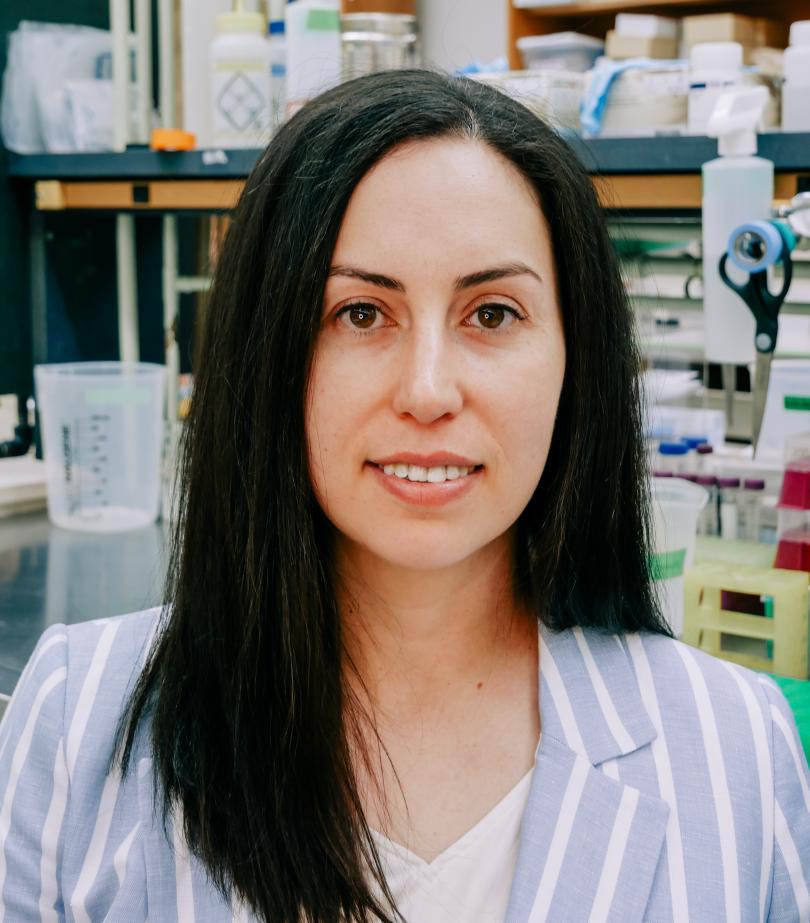
Dr. Deb Bell-Pedersen smiling at the camera. She is wearing glasses and hanging earrings.Dr. Deb Bell-Pedersen | Pedersen Lab

Abstract:
The circadian clock is a fundamental regulator of human health and drug metabolism, coordinating daily rhythms in protein production that affect cellular function and metabolism. Many proteins that cycle robustly are produced from non-rhythmic mRNAs, pointing to translational control as a key mechanism of rhythmic protein levels. Using the model eukaryote Neurospora crassa, we discovered that the clock exerts this regulation through rhythmic control of a conserved translation initiation factor (eIF2α) and by remodeling ribosome composition. Even more unexpectedly, we discovered that the circadian system governs the fidelity of protein synthesis by modulating ribosome makeup and tRNA synthetase activity. Both translational fidelity and circadian amplitude decline with age. We identified compounds that restore clock amplitude in old N. crassa cells, leading to improved translation accuracy and extended lifespan. These findings reveal how the circadian clock programs daily changes in the proteome beyond genomic instructions and highlight a novel link between circadian regulation, proteome integrity, and aging.

Date:
Location:
THM 116

"Understanding the Sources of Regenerative Capacity in Animals"

Dr. Alejandro Sánchez Alvarado standing in front of a white dry erase board with his arms crossed while smiling. The dry erase board behind him has biology graphics in different colors.

Dr. Alejandro Sánchez Alvarado | Sánchez Alvarado Lab

Bio:
Sánchez Alvarado received a BS in molecular biology and chemistry from Vanderbilt University in Nashville, TN, and a PhD in pharmacology and cell biophysics from the University of Cincinnati College of Medicine in Cincinnati, OH. He performed postdoctoral and independent research at the Carnegie Institution of Washington, Department of Embryology in Baltimore, MD. In 2002, he joined the faculty of the University of Utah School of Medicine in Salt Lake City where he held the H.A. & Edna Benning Presidential Endowed Chair. In 2005, he was named a Howard Hughes Medical Institute Investigator. He joined the Stowers Institute for Medical Research in Kansas City in 2011 and became the President and Chief Scientific Officer of the Stowers Institute in 2022. He also holds the Priscilla Wood Neaves Chair in the Biomedical Sciences.

Sánchez Alvarado is an elected member of the National Academy of Science, the American Academy of Arts and Sciences, and the Latin American Academy of Sciences, a Kavli Fellow of the National Academy of Sciences USA, a Fellow of the Marine Biological Laboratory in Woods Hole, MA, a Fellow of the American Association for the Advancement of Science, and a recipient of a National Institutes of Health MERIT award, the EE Just Medal for Scientific Achievement, and the Vilcek Prize in Biomedical Sciences. He has served on numerous scientific advisory committees and boards including the National Advisory Council of the National Institute of General Medical Sciences, National Institutes of Health, and presently serves on the Board of Directors of American Century Investments.

Sánchez Alvarado’s work has the potential to lead to a better understanding of how the adult forms of higher organisms, including humans, carry out their biological functions. His research also has led to insights on the molecular and genetic drivers of both regenerative and degenerative cellular processes that contribute to disease.

Abstract:
It is paradoxical that for many organisms (including humans), the apparent anatomical stability of their adult bodies is maintained by constant change. Despite the importance of tissue homeostasis and regeneration to human biology and health, relatively little is known about how these processes are regulated. As such, numerous questions remain unanswered, including: How do organ systems maintain their order and function while in a state of cell flux? How do animals control and coordinate the size and cell number of multiple organ systems? Does regeneration of body parts lost to injury invoke embryonic processes, generic patterning mechanisms, or unique circuitry comprised of well-established patterning genes? Answering any of these questions would set a baseline from which to try to enhance regenerative properties in multicellular organisms such as humans, particularly after injury.

One way to solve a complex problem is to reduce it to a simpler, easier to answer problem. Therefore, reducing the complexities of regeneration and tissue homeostasis to the study of comparatively simpler systems would allow for a systematic dissection and mechanistic understanding of these processes. Here, I will discuss how the use of single-cell and spatial transcriptomics is helping define the cellular and molecular environments that support pluripotency in the highly regenerative freshwater planarian Schmidtea mediterranea and regeneration of missing organs in the hemichordate Ptychodera flava. Our studies are beginning to shed light on the way adult animals regulate tissue homeostasis and the replacement of body parts lost to injury.

Date:
Location:
THM 116

"An Active Role for Vision Prior to Eye-opening in Neonates"

Photo of Dr. D'Souza smiling in a white lab coat. In the background you can see blurred green leaves from a tree in the back right corner and the side of a building in the back left corner.Dr. Shane D'Souza

Bio:
Dr. Shane Peter D’Souza is a neuroscientist and vision researcher whose work spans developmental neurobiology, sensory physiology, and circadian biology. He earned his BS in Biology at the University of Kentucky and PhD in Molecular and Developmental Biology from the University of Cincinnati/Cincinnati Children’s Hospital Medical Center, where he investigated how early light exposure shapes neural circuit development in the retina and brain. Now a Postdoctoral Research Fellow in Pediatric Ophthalmology at Cincinnati Children’s, Dr. D’Souza’s research integrates molecular, anatomical, physiological, and computational approaches to understand how intrinsically photosensitive retinal ganglion cells (ipRGCs) and other non-visual photoreceptors influence sensory-driven circuit refinement, photoreceptor abundance, and cross-modal communication in early life. His publication record includes discoveries on melanopsin-dependent regulation of rod photoreceptors, neuropsin and encephalopsin function in visual and non-visual systems, and the role of light-sensitive hypothalamic neurons in thermoregulation and metabolism. By combining high-resolution imaging, transcriptomics, biophysical modeling, and machine-learning, his work aims to uncover fundamental principles of sensory-mediated neural development and sensory adaptation across species. In his spare time, he enjoys studying the evolution of storm systems and serves as a storm spotter for NWS Wilmington, OH. In his spare-spare time, he writes and produces music from his living room.

Abstract:
Most mammals are born with immature, poorly developed sensory systems. As these systems come online, they use immature sensory experience to shape synapses, cell types, and their connectivity across the brain. In the visual system, this experience is thought to be passive, supporting and setting up later modes of image-forming vision after eye-opening.  However, little is known about the form and function of visual experience during the earliest period in a neonate’s life. Driven by this, we set out to generate a comprehensive map of visual system activity and neonatal behaviors in mice.  Using a host of machine learning-based approaches we developed an atlas perinatal visual system activity from the retina to several regions of the brain. Using a combination of chromatic stimuli and genetic loss-of-function mice, we identified the M1 intrinsically photosensitive retinal ganglion cell (ipRGC) in the retina as the driver of early visual system activity, activating distinct brain regions during development. Using this atlas as a guide to assess behaviors, we find that this visual input drives the production of ultrasonic vocalization in neonates and “blinding” mice leads to an augmented vocal code.  Together, these data suggest that early visual system activity has an active role in supporting the development of neonatal behaviors and warrants a deeper exploration of early sensory activity across the developing brain.

A photo of Dr. D'Souza in a white lab coat crouched in some grass holding a doggie toy. Across from Dr. D'Souza is a Golden Retriever with a harness on and it's tongue hanging out of its mouth.Pictured is a color enhanced mouse brain.


Date:
Location:
THM 116