
"Fishing out the Function of Structural and Receptor Proteins in Photoreceptors and the Retinal Pigmented Epithelium"
Date:
-
Location:
THM 116
Event Series:

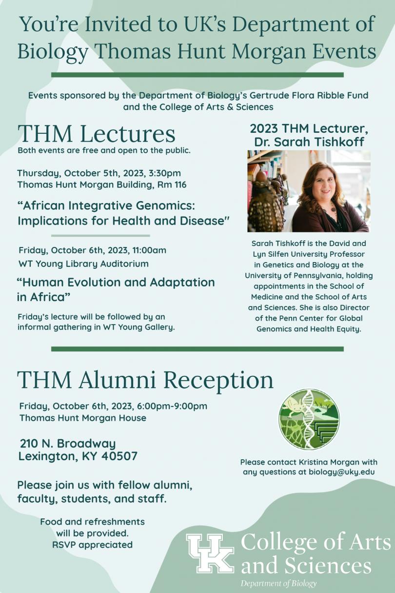
Click here for more information about Dr. Sarah Tishkoff.
Abstract:
Africa is thought to be the ancestral homeland of all modern human populations. It is also a region of tremendous cultural, linguistic, climatic, and genetic diversity. Despite the important role that African populations have played in human history, they remain one of the most underrepresented groups in human genomics studies. A comprehensive knowledge of patterns of variation in African genomes is critical for a deeper understanding of human genomic diversity, the identification of functionally important genetic variation, the genetic basis of adaptation to diverse environments and diets, and for reconstructing modern human origins. African populations practice diverse subsistence patterns (hunter-gatherers, pastoralists, agriculturalists, and agro-pastoralists) and live in diverse environments with differing pathogen exposure (tropical forest, savannah, coastal, desert, low altitude, and high altitude) and, therefore, are likely to have experienced local adaptation. In this talk I will discuss results of analyses of genome-scale genetic variation in geographically, linguistically, and ethnically diverse African populations in order to reconstruct human evolutionary history in Africa, African and African American ancestry, as well as the genetic basis of adaption to diverse environments.