
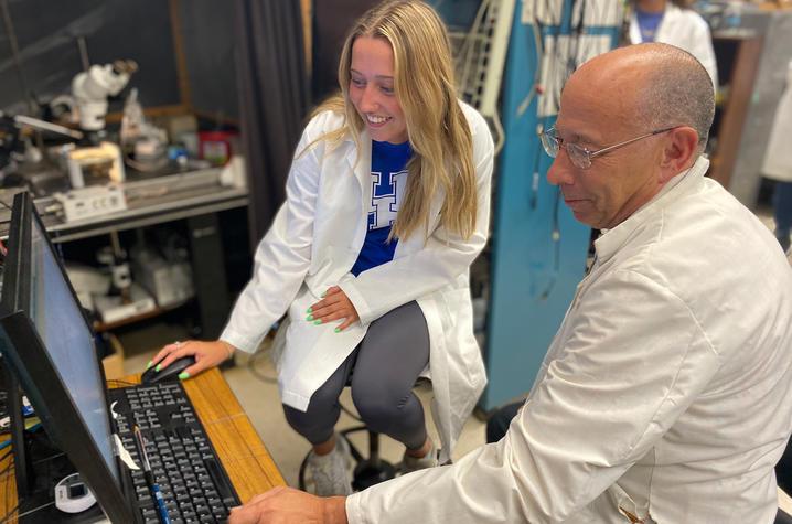
Biology alum's research could have eye-opening implications for NICU; his journey started here.
By Francis Von Mann
LEXINGTON, Ky. (Oct. 8, 2025) – In the Don & Cathy Jacobs Science Building, a mural sprawls across a whiteboard. On one side, a magnified cell with swirling, intricate layers. On the other, Charles Darwin circled by Galápagos finches and double helixes. It is equal parts science and art, memorialized with a signature in the corner: Shane D’Souza, Class of 2018.
Faculty Emeritus


Master's Student


Teaching Postdoctoral Scholar
Graduate Student

Cognitive Neuroscience Graduate Student

Teaching Assistant, French & Francophone Studies

Department Manager
Professor Emeritus


Senior Lecturer
Graduate Student and Teaching Assistant, Biology

PhD candidate

Postdoctoral Scholar

Otis A. Singletary Chair in the Humanities


Environmental Scientist

Associate Professor, Linguistics

MFA Candidate
Part Time Instructor

Senior Research Geologist, Kentucky Geological Survey

Graduate Student

Academic Advisor II


Director Louie B. Nunn Center for Oral History

Assistant Dean for Communications and Technical Solutions

Associate Professor


Professor

Graduate Research Assistant

Lecturer

Academic Advisor

Neuroscience

Professor
"Mom does it best: Parental Care as a Model Phenotype to Explore How Cell-Type Specific Changes in Gene Expression Influence Brain Activity and Animal Behavior"
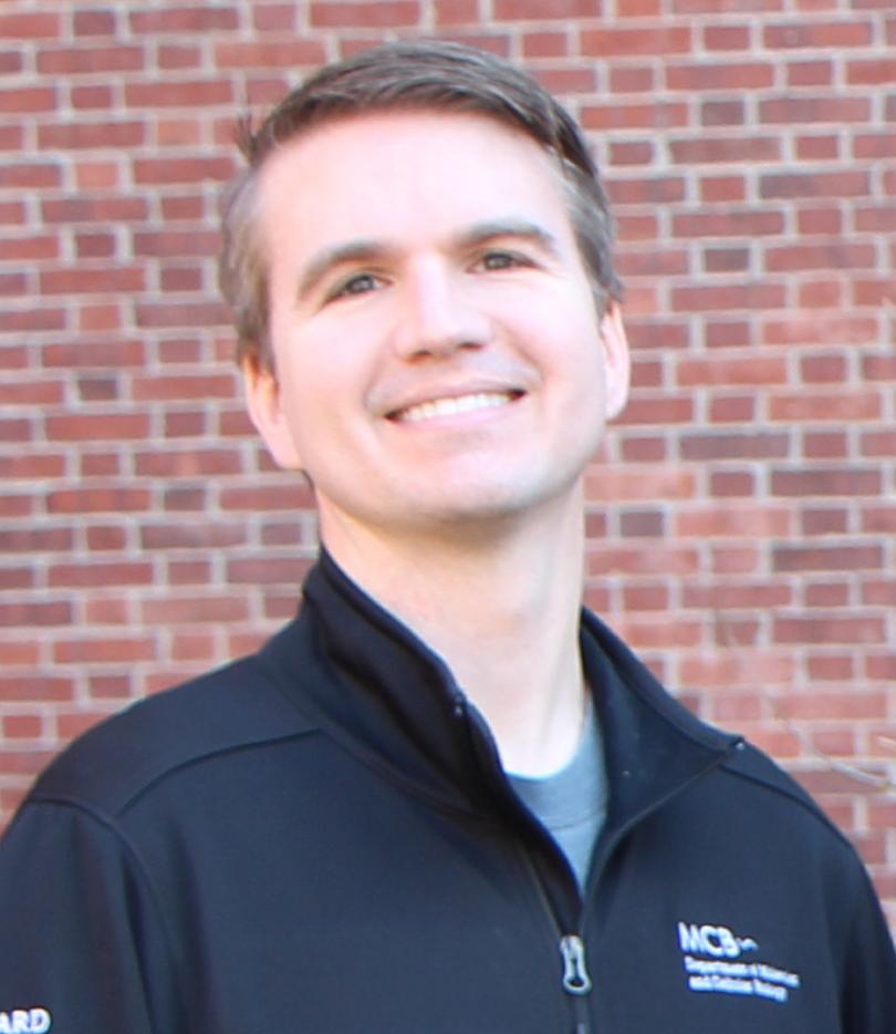 Dr. Brandon Logeman
Bio:
Brandon L. Logeman, PhD is a new Assistant Professor in the Department of Molecular and Cellular Biochemistry, College of Medicine, University of Kentucky. After completing his Ph.D. at Duke University, he joined the lab of Catherine Dulac at Harvard University to study the molecular mechanisms through which changes in cell-type specific gene expression influence neural activity and animal behavior. After receiving a K99/R00 Career Transition Award he joined the University of Kentucky in August 2025. His new lab will utilize custom designed single-cell genomics technologies such as microfluidic, droplet based sequencing assays and imaging based spatial transcriptomics as well as de novo protein binder design across a panel of genetically diverse mouse strains to discover how genomic and environmental influences contribute to observable differences in animal behavior.
Abstract:
Parental care is composed of multiple infant-directed behaviors that promote offspring survival and is influenced by the sex and physiological state of the caregiver. Previous work in mice has identified the medial preoptic area of the hypothalamus as a key brain area implicated in parental behaviors. However, numerous naturalistic behaviors and homeostatic processes are controlled by this area, hindering mechanistic investigation of the circuits underlying parental care. To overcome this challenge, here I employ cell-type specific RNA- and ATAC-seq analysis, neural activity recording, and perturbation to gain access into molecular, biophysical, and circuit-based causality of behavioral control. I find that various neuronal types involved in parenting behavior are each distinctively influenced by the sex and physiological status of an individual and uncover how cell-type specific regulatory programs alter gene expression and neural activity underlying behavior control. These results demonstrate how cell-type specific transcriptional responses to internal physiological cues mediate circuit specific alterations to neural activity and ultimately influence animal behavior.
Date:
Location:
THM 116
Event Series:
'From Prokaryotes to Superorganisms: Constraints on the Evolution and Distribution of Size, Diversity, and Complexity'
 Dr. Jordan Okie | Okie Additional Information
Biosketch:
Jordan Okie is a research professor in the School of Earth and Space Exploration at Arizona State University. Motivated by a variety of field experiences spanning the extremes of life and its environments, his research aims to understand patterns of biodiversity, scaling, metabolism and macroevolution across levels of organization, from tiny prokaryotes to giant multicellular organisms, ecosystems, colonies and technological systems. His research combines mathematical theory and modeling, bioinformatics, microbial experiments and the compilation and analysis of comparative data sets. He is also concerned with issues of Anthropocene ecology and sustainability, authoring a book and widely-cited papers on these topics. Recent notable papers include work on the rarity of prokaryote endosymbioses, genomic adaptations in whole-ecosystem experiments and the development of the Equilibrium Theory of Biodiversity Dynamics.
He earned a Ph.D. in biology from the University of New Mexico in 2011 and has held fellowships and visiting appointments with NASA’s Astrobiology Institute, the Czech Science Foundation’s Center for Theoretical Study and the Santa Fe Institute. He joined the faculty at ASU in 2015 and teaches courses on astrobiology, biogeochemistry and ecology.
Abstract:
The diversity, size and complexity of living things on Earth today vary widely across the globe, following some general patterns of distribution whose underlying mechanisms remain hotly contested. The evolution of the largest and most complex living things required some 3.8 billion years and massive amounts of energy.
This seminar will explore the general ecological, evolutionary, physiological and genomic factors limiting and shaping this extraordinary biological variation across space and time. The seminar will present theory, experiments and comparative analyses revealing the role of biological innovations, including evolutionary transitions in individuality in facilitating the evolution of larger-bodied living things.
The seminar will cover work on metabolic scaling, metabolic modeling, metagenomics and biodiversity theory that sheds light on the role of energetic constraints in shaping the distribution and evolution of the biodiversity, body size and hierarchical complexity of living things, from prokaryotes to single-celled eukaryotes, complex endosymbioses, multicellular organisms, holobionts, superorganisms and technological systems.
Watch the seminar here.
Date:
Location:
THM 116
Event Series:
"Circadian Rhythms in Hosts, Parasites and their Vectors"
 Dr. Filipa Rijo-Ferreira | Rijo-Ferreira Lab
Bio:
Rijo-Ferreira is an assistant professor of public health and molecular and cellular biology at the University of California, Berkeley, and an investigator at the Chan Zuckerberg BioHub. Rijo-Ferreira earned a Ph.D. from University of Porto, Portugal, followed by postdoctoral training at the University of Texas Southwestern Medical Center at Dallas. The Rijo-Ferreira lab takes an integrated approach to study circadian rhythms in parasitic diseases, in particular Malaria and Sleeping sickness. Rijo-Ferreira is an NIH Pathway to Independence awardee, a NIH Director’s New Innovator awardee and a Searle scholar. Moreover, Rijo-Ferreira was recently named a HHMI Freeman Hrabowski Scholar, which recognizes outstanding early career faculty for their research and commitment to mentoring.
Date:
Location:
THM 116
Event Series:
'Ecological Responses Aren’t Instantaneous, and That’s Important: Rate-dependent Tipping and Transients'
 Dr. Karen Abbott | Abbott Lab
Bio:
Dr. Karen Abbott is a theoretical ecologist in the Department of Biology at Case Western Reserve University. Her research spans a range of topics with an emphasis on general principles that underly many ecological mechanisms and systems. She received her B.S. in biology and mathematics at Vanderbilt University, and a Ph.D. in ecology & evolution from the University of Chicago.
Abstract:
Rate dependent tipping occurs when external conditions change too quickly for the ecological dynamics to track. Long transients occur when ecological dynamics are slow to equilibrate. In both cases, the root cause is that ecological systems don’t respond instantaneously to changes. To what extent are transients and rate dependence different sides of the same coin? And, can classical theoretical ecology, with its strong focus on equilibria, be at all helpful for understanding these phenomena? In this talk, I will discuss how our understanding of equilibria helps us understand transients and how I think our understanding of transients may be a fruitful approach to understanding rate-dependent tipping.
Date:
Location:
THM 116
Event Series:
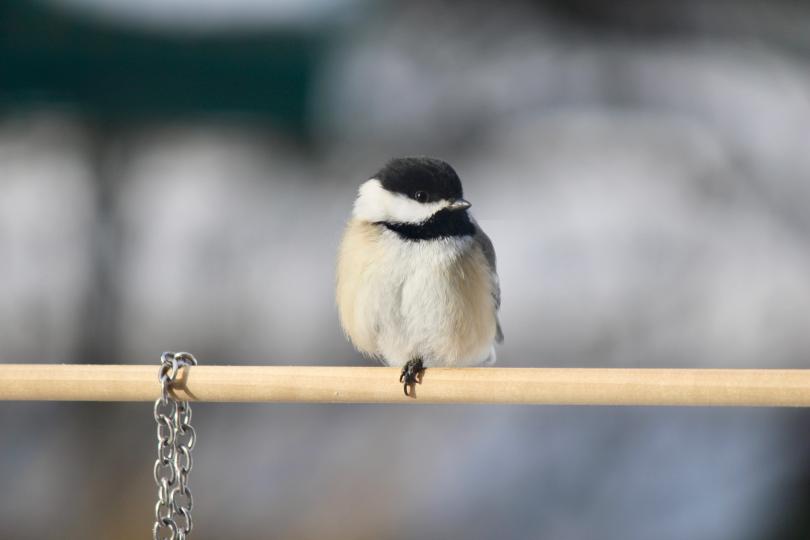
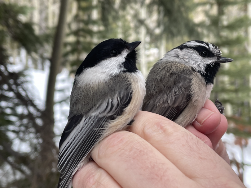
"Hybridization and Adaptation in Chickadees"

Dr. Scott Taylor | Taylor Lab
Bio:
Dr. Scott Taylor is the director of the CU Boulder Mountain Research Station. Research in his group focuses on using natural hybrid zones and recent radiations to understand the genetic bases of traits involved in reproductive isolation, population divergence and speciatio, and the impacts of anthropogenic change, including climate change, on species distributions, interactions and evolution. We're fascinated by natural history and the intersections between art and science, and we're committed to doing our part to support our community.
Abstract:
Chickadees are familiar and widespread nonmigratory birds that have amazing adaptations to survive cold winters, including the ability to remember hundreds of thousands of individual seed cache locations. Recovering these caches is critical for winter survival. Taylor will share recent results from two different studies, one focused on a chickadee hybrid zone and one focused on a single population of mountain chickadees, that both provide insights into the genetic basis of variation in spatial cognition in chickadees.
Date:
Location:
THM 116
Event Series: